
The doctor is coming,
what should you recommend to help the patient?
A great start!
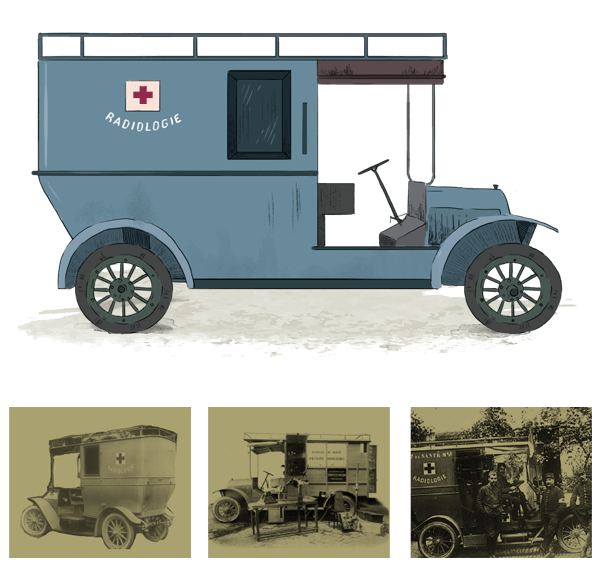
X-rays were discovered in 1895 and physicians started using them shortly after. X-ray machines were only in big city hospitals at the beginning of the war and weren't very useful in the war effort. But celebrated scientist Marie Curie invented a "radiological car" that brought x-rays to the front. Though she trained over 150 women to operate these cars, x-rays weren't available in every field hospital.
But bravo, you've managed to get an x-ray for your patient and the femur is broken!
What now?
Great instinct!
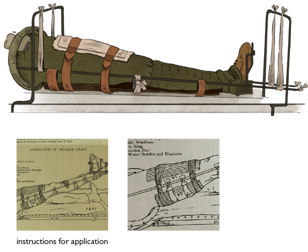
The right kind of splint is perfect to immobilize the bone, while ensuring that the open wound doesn't get infected. It will also ensure comfort as the patient is transported, since they'll have to be moved away from the front for a long recovery.
The "Thomas Splint," though first used in 1865, only came into widespread use in WWI. Fractures of the femur were very dangerous in WWI. Before the Thomas Splint was used, fractured femurs had a mortality rate of 80% due to internal bleeding or infection. The Thomas Splint reduced that rate to 20%!
It looks very complicated but it's actually still used today, particularly in military settings.

Not quite!
Though plaster casts were in use in WWI and were vastly improved during the war (notably by Irish sculpture Anne Acheson) a plaster cast is not the best choice for a femur fracture, which had an extremely high fatality rate in WWI, especially with the presence of an open wound.
Close